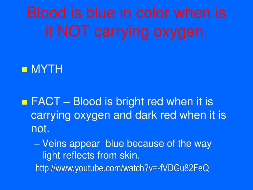
Why Veins Look Blue Through Skin Even Though Human Blood Is Always Red
The mystery is physical, not biochemical. Sunlight and artificial ambient lighting consist of broad white light, spanning wavelengths from approximately 380 nm (violet) to 750 nm (deep red).
When white light strikes human skin, photons do not bounce off a single surface like a mirror. Instead, they penetrate multiple layers of tissue: the epidermis, the dermis, and the hypodermis.
Blue light has shorter wavelengths (around 400, 495 nm) and carries high scattering energy. It cannot penetrate deeply into human tissue; it scatters rapidly within the upper layers of the dermis and bounces back out toward the viewer.
Red light has longer wavelengths (around 620, 750 nm) and penetrates much deeper into tissue, often reaching 5 to 10 millimeters below the skin surface.
| Wavelength Band | Typical Depth in Dermis | Hemoglobin Absorption | Perceived Surface Result |
|---|---|---|---|
| Blue (400, 495 nm) | 0.5, 1.0 mm | Low (scattered before vessel depth) | Reflected back to the eye |
| Green (495, 570 nm) | 1.0, 2.5 mm | Moderate to high | Largely extinguished in upper dermis |
| Red (620, 750 nm) | 5.0, 10.0 mm | Extremely high absorption by blood | Absorbed by vein; background skin reflects red |
Most subcutaneous veins sit approximately 1 to 2 millimeters beneath the surface. When red light penetrates that deep, it strikes the dark red blood within the vessel. Hemoglobin absorbs red photons heavily.
Because the vein absorbs red light, less red light reflects back from the precise area above the vessel compared to the surrounding skin. Meanwhile, blue light scatters out from the superficial dermis across the entire arm.
The human visual cortex processes this contrast dynamically. Next to the reddish reflection of surrounding tissue, the localized deficit of red light directly above the vessel causes the brain to perceive the vein as blue.